Causes Of Pain In The Foot Arch
Overview
The most common causes of arch pain are repetitive activities, such as walking or running too far; jumping; or standing on hard surfaces for long periods of time. Injury, such as tears in the ligament called the plantar fascia. The natural aging process. Biomechanical defects of the foot, or abnormalities we are born with, such as very high or low arches, or pronation (a turning out of the foot, causing us to walk more on the inner border of the foot). Plantar fibromatosis, a benign growth on the ligament that supports the arch.

Causes
The arches are the primary structures of the body that absorb and return force to and from the body to the outside world when we are on our feet. When something happens to these structures, pain and injury may result. There can be many causes of arch pain. Direct force trauma, ligament sprains, muscle strains, poor biomechanical alignment,stress fractures, overuse, inflammatory arthritis or the tightness or lack of tightness of the joints in the foot may all cause pain in the arch. Injury to the plantar fascia is a common cause of arch pain. The plantar fascia is the thick, connective tissue which supports the arch on the bottom of the foot. It runs from the calcaneus (heel) forwards to the heads of the metatarsals. When the plantar fascia is damaged, the resulting inflammatory response may become a source of arch pain.
Symptoms
Flat feet can exhibit a variety of symptoms, from mild to severe. The extent of the flat foto does not always correlate with the extent of symptoms. Patients may complain of arch pain and heel pain. Commonly there is pain on the outside of the foot, where the foot meets the ankle as the collapse foot abuts against the ankle. Muscle cramps within the foot, and onto the leg (shin splints) may occur. In general, patients have pain with activity, such as walking or running. The pain may be deep and focal to a generalized widespread achy feeling. Irritation from shoe gear can cause redness and swelling. Common reasons patients seek treatment are pain, interference with walking or activities, difficulty fitting shoes, swelling, and notice a change in appearance of the foot and/or unsightly appearance.
Diagnosis
The medical practitioner will examine how the muscles of your foot function. These tests may involve holding or moving your foot and ankle against resistance; you may also asked to stand, walk, or even run. Pain caused by movements may indicate the cause of the pain. The nerves in the foot will be tested to make sure no injury has occurred there. An x-ray, MRI, or bone scan of the foot and arch may be taken to determine if there are changes in the makeup of the bone.
Non Surgical Treatment
How the pain in the bottom of your foot is treated will depend heavily on the cause of the pain. Diagnosing the pain while it?s in the early stages is important when determining the best treatment options. If the pain is mild to moderate, simple improvements in footwear can help reduce the symptoms. Most patients must use the RICE method for effective treatment. RICE stands for Rest, Ice, Compression, and Elevation. This is a popular treatment used by athletes. It involves resting the foot, icing it for fifteen to twenty minute intervals, compressing the foot with a bandage, and elevating it at least twelve inches above the heart. Ant-inflammatory and pain medications are also sometimes used to treat bottom-of-foot pain. For more serious cases, steroid injections or foot surgery may help reduce pain and swelling and correct the underlying condition (if there is one.) If you suffer from a severe case of plantar fasciitis and non-surgical methods fail, your doctor may recommend cortisone injections to relieve the pain. If cortisone injections fail, your doctor may recommend a surgical procedure that involves cutting and releasing the plantar fascia.

Surgical Treatment
Surgical advances have dramatically improved the ability to alleviate the pain and decreased function that millions of Americans experience due to flat feet. Nevertheless, many patients and even some physicians remain unaware of the new procedures, which are best performed by a foot and ankle specialist who has the applicable training and experience.
Prevention
Maintain a healthy weight, Use insoles to support your arches, Limit how often you wear high heels, Use proper shoes, especially when exercising to evenly distribute weight through your foot.
The most common causes of arch pain are repetitive activities, such as walking or running too far; jumping; or standing on hard surfaces for long periods of time. Injury, such as tears in the ligament called the plantar fascia. The natural aging process. Biomechanical defects of the foot, or abnormalities we are born with, such as very high or low arches, or pronation (a turning out of the foot, causing us to walk more on the inner border of the foot). Plantar fibromatosis, a benign growth on the ligament that supports the arch.
Causes
The arches are the primary structures of the body that absorb and return force to and from the body to the outside world when we are on our feet. When something happens to these structures, pain and injury may result. There can be many causes of arch pain. Direct force trauma, ligament sprains, muscle strains, poor biomechanical alignment,stress fractures, overuse, inflammatory arthritis or the tightness or lack of tightness of the joints in the foot may all cause pain in the arch. Injury to the plantar fascia is a common cause of arch pain. The plantar fascia is the thick, connective tissue which supports the arch on the bottom of the foot. It runs from the calcaneus (heel) forwards to the heads of the metatarsals. When the plantar fascia is damaged, the resulting inflammatory response may become a source of arch pain.
Symptoms
Flat feet can exhibit a variety of symptoms, from mild to severe. The extent of the flat foto does not always correlate with the extent of symptoms. Patients may complain of arch pain and heel pain. Commonly there is pain on the outside of the foot, where the foot meets the ankle as the collapse foot abuts against the ankle. Muscle cramps within the foot, and onto the leg (shin splints) may occur. In general, patients have pain with activity, such as walking or running. The pain may be deep and focal to a generalized widespread achy feeling. Irritation from shoe gear can cause redness and swelling. Common reasons patients seek treatment are pain, interference with walking or activities, difficulty fitting shoes, swelling, and notice a change in appearance of the foot and/or unsightly appearance.
Diagnosis
The medical practitioner will examine how the muscles of your foot function. These tests may involve holding or moving your foot and ankle against resistance; you may also asked to stand, walk, or even run. Pain caused by movements may indicate the cause of the pain. The nerves in the foot will be tested to make sure no injury has occurred there. An x-ray, MRI, or bone scan of the foot and arch may be taken to determine if there are changes in the makeup of the bone.
Non Surgical Treatment
How the pain in the bottom of your foot is treated will depend heavily on the cause of the pain. Diagnosing the pain while it?s in the early stages is important when determining the best treatment options. If the pain is mild to moderate, simple improvements in footwear can help reduce the symptoms. Most patients must use the RICE method for effective treatment. RICE stands for Rest, Ice, Compression, and Elevation. This is a popular treatment used by athletes. It involves resting the foot, icing it for fifteen to twenty minute intervals, compressing the foot with a bandage, and elevating it at least twelve inches above the heart. Ant-inflammatory and pain medications are also sometimes used to treat bottom-of-foot pain. For more serious cases, steroid injections or foot surgery may help reduce pain and swelling and correct the underlying condition (if there is one.) If you suffer from a severe case of plantar fasciitis and non-surgical methods fail, your doctor may recommend cortisone injections to relieve the pain. If cortisone injections fail, your doctor may recommend a surgical procedure that involves cutting and releasing the plantar fascia.
Surgical Treatment
Surgical advances have dramatically improved the ability to alleviate the pain and decreased function that millions of Americans experience due to flat feet. Nevertheless, many patients and even some physicians remain unaware of the new procedures, which are best performed by a foot and ankle specialist who has the applicable training and experience.
Prevention
Maintain a healthy weight, Use insoles to support your arches, Limit how often you wear high heels, Use proper shoes, especially when exercising to evenly distribute weight through your foot.
Achilles Tendonitis Taping
Overview
 Achilles tendinitis is very common among running athletes. The calf muscles attach to the calcaneus via the Achilles tendon. During running, the calf muscles help with the lift-off phase of gait. Repetitive forces from running combined with insufficient recovery time can initially cause inflammation in the tendon paratenon (fatty areolar tissue that surrounds the tendon). A complete tear of the Achilles tendon is a serious injury, usually resulting from sudden, forceful stress. Tendon tears can occur with minimal exertion in people who have taken fluoroquinolone antibiotics.
Causes
Achilles tendonitis is a common sports injury caused by repeated or intense strain on the tendon. But non-athletes also can get it if they put a lot of stress on their feet. Other things that contribute to Achilles tendonitis include. An increase in activity. Starting a training program after a period of inactivity or adding miles or hills to a jogging regimen are two examples of things that put people at risk for Achilles tendonitis. Sports that require sudden starts and stops; for example, tennis and basketball. A change in footwear, or wearing old or badly fitting shoes. New shoes, worn-out shoes, or the wrong size shoes can cause a person's feet to overcompensate and put stress on the Achilles tendon. Additionally, wearing high heels all the time can cause the tendon and calf muscles to get shorter, and the switch to flat shoes and exercise can put extra strain on the heel. Running up hills. Going uphill forces the Achilles tendon to stretch beyond its normal range. Weak calf muscles, flat arches, "overpronation" (feet that roll in when running), or "oversupination" (feet that roll out when running). Overpronation and oversupination make the lower leg rotate and put a twisting stress on the tendon. Exercising without warming up. Tight calf muscles or muscles that lack flexibility decrease a person's range of motion and put an extra strain on the tendon. Running or exercising on a hard or uneven surface or doing lunges or plyometrics without adequate training. A traumatic injury to the Achilles tendon.
Symptoms
Symptoms of Achilles tendinitis and tendinosis include recurring localized heel pain, sometimes severe, along the achilles tendon during or after exercise. Pain often begins after exercise and gradually worsens. Morning tenderness or stiffness about an inch and a half above the point where the Achilles tendon is attached to the heel bone. Sluggishness in your leg. Mild to severe swelling. Stiffness that generally diminishes as the tendon warms up with use.
Diagnosis
Physicians usually pinch your Achilles tendon with their fingers to test for swelling and pain. If the tendon itself is inflamed, your physician may be able to feel warmth and swelling around the tissue, or, in chronic cases, lumps of scar tissue. You will probably be asked to walk around the exam room so your physician can examine your stride. To check for complete rupture of the tendon, your physician may perform the Thompson test. Your physician squeezes your calf; if your Achilles is not torn, the foot will point downward. If your Achilles is torn, the foot will remain in the same position. Should your physician require a closer look, these imaging tests may be performed. X-rays taken from different angles may be used to rule out other problems, such as ankle fractures. MRI (magnetic resonance imaging) uses magnetic waves to create pictures of your ankle that let physicians more clearly look at the tendons surrounding your ankle joint.
Nonsurgical Treatment
NSAIDS like ibuprofen are often prescribed to help manage the pain and inflammation. Steroids are often recommended when patients do not respond to NSAID treatment. They are often most effective when injected directly into the inflamed and swollen area. Physiotherapy is a great way to stretch and strengthen the Achilles tendon. A good physical therapist will also teach the patient techniques which give better foot support during exercise (taping, wrapping, etc?). Orthotics, assistive devices and insoles can be used to cushion and cradle the arch of the foot during the healing process. Shock Wave Therapy. This is the newest form of treatment and uses concentrated sound waves to stimulate healing in the affected area. This form of treatment is reserved for heel pain that is unresponsive to other forms of treatment.

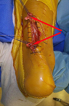
Surgical Treatment
If several months of more-conservative treatments don't work or if the tendon has torn, your doctor may suggest surgery to repair your Achilles tendon.
Prevention
While it may not be possible to prevent Achilles tendinitis, you can take measures to reduce your risk. Increase your activity level gradually. If you're just beginning an exercise regimen, start slowly and gradually increase the duration and intensity of the training. Take it easy. Avoid activities that place excessive stress on your tendons, such as hill running. If you participate in a strenuous activity, warm up first by exercising at a slower pace. If you notice pain during a particular exercise, stop and rest. Choose your shoes carefully. The shoes you wear while exercising should provide adequate cushioning for your heel and should have a firm arch support to help reduce the tension in the Achilles tendon. Replace your worn-out shoes. If your shoes are in good condition but don't support your feet, try arch supports in both shoes. Stretch daily. Take the time to stretch your calf muscles and Achilles tendon in the morning, before exercise and after exercise to maintain flexibility. This is especially important to avoid a recurrence of Achilles tendinitis. Strengthen your calf muscles. Strong calf muscles enable the calf and Achilles tendon to better handle the stresses they encounter with activity and exercise. Cross-train. Alternate high-impact activities, such as running and jumping, with low-impact activities, such as cycling and swimming.
Achilles tendinitis is very common among running athletes. The calf muscles attach to the calcaneus via the Achilles tendon. During running, the calf muscles help with the lift-off phase of gait. Repetitive forces from running combined with insufficient recovery time can initially cause inflammation in the tendon paratenon (fatty areolar tissue that surrounds the tendon). A complete tear of the Achilles tendon is a serious injury, usually resulting from sudden, forceful stress. Tendon tears can occur with minimal exertion in people who have taken fluoroquinolone antibiotics.
Causes
Achilles tendonitis is a common sports injury caused by repeated or intense strain on the tendon. But non-athletes also can get it if they put a lot of stress on their feet. Other things that contribute to Achilles tendonitis include. An increase in activity. Starting a training program after a period of inactivity or adding miles or hills to a jogging regimen are two examples of things that put people at risk for Achilles tendonitis. Sports that require sudden starts and stops; for example, tennis and basketball. A change in footwear, or wearing old or badly fitting shoes. New shoes, worn-out shoes, or the wrong size shoes can cause a person's feet to overcompensate and put stress on the Achilles tendon. Additionally, wearing high heels all the time can cause the tendon and calf muscles to get shorter, and the switch to flat shoes and exercise can put extra strain on the heel. Running up hills. Going uphill forces the Achilles tendon to stretch beyond its normal range. Weak calf muscles, flat arches, "overpronation" (feet that roll in when running), or "oversupination" (feet that roll out when running). Overpronation and oversupination make the lower leg rotate and put a twisting stress on the tendon. Exercising without warming up. Tight calf muscles or muscles that lack flexibility decrease a person's range of motion and put an extra strain on the tendon. Running or exercising on a hard or uneven surface or doing lunges or plyometrics without adequate training. A traumatic injury to the Achilles tendon.
Symptoms
Symptoms of Achilles tendinitis and tendinosis include recurring localized heel pain, sometimes severe, along the achilles tendon during or after exercise. Pain often begins after exercise and gradually worsens. Morning tenderness or stiffness about an inch and a half above the point where the Achilles tendon is attached to the heel bone. Sluggishness in your leg. Mild to severe swelling. Stiffness that generally diminishes as the tendon warms up with use.
Diagnosis
Physicians usually pinch your Achilles tendon with their fingers to test for swelling and pain. If the tendon itself is inflamed, your physician may be able to feel warmth and swelling around the tissue, or, in chronic cases, lumps of scar tissue. You will probably be asked to walk around the exam room so your physician can examine your stride. To check for complete rupture of the tendon, your physician may perform the Thompson test. Your physician squeezes your calf; if your Achilles is not torn, the foot will point downward. If your Achilles is torn, the foot will remain in the same position. Should your physician require a closer look, these imaging tests may be performed. X-rays taken from different angles may be used to rule out other problems, such as ankle fractures. MRI (magnetic resonance imaging) uses magnetic waves to create pictures of your ankle that let physicians more clearly look at the tendons surrounding your ankle joint.
Nonsurgical Treatment
NSAIDS like ibuprofen are often prescribed to help manage the pain and inflammation. Steroids are often recommended when patients do not respond to NSAID treatment. They are often most effective when injected directly into the inflamed and swollen area. Physiotherapy is a great way to stretch and strengthen the Achilles tendon. A good physical therapist will also teach the patient techniques which give better foot support during exercise (taping, wrapping, etc?). Orthotics, assistive devices and insoles can be used to cushion and cradle the arch of the foot during the healing process. Shock Wave Therapy. This is the newest form of treatment and uses concentrated sound waves to stimulate healing in the affected area. This form of treatment is reserved for heel pain that is unresponsive to other forms of treatment.
Surgical Treatment
If several months of more-conservative treatments don't work or if the tendon has torn, your doctor may suggest surgery to repair your Achilles tendon.
Prevention
While it may not be possible to prevent Achilles tendinitis, you can take measures to reduce your risk. Increase your activity level gradually. If you're just beginning an exercise regimen, start slowly and gradually increase the duration and intensity of the training. Take it easy. Avoid activities that place excessive stress on your tendons, such as hill running. If you participate in a strenuous activity, warm up first by exercising at a slower pace. If you notice pain during a particular exercise, stop and rest. Choose your shoes carefully. The shoes you wear while exercising should provide adequate cushioning for your heel and should have a firm arch support to help reduce the tension in the Achilles tendon. Replace your worn-out shoes. If your shoes are in good condition but don't support your feet, try arch supports in both shoes. Stretch daily. Take the time to stretch your calf muscles and Achilles tendon in the morning, before exercise and after exercise to maintain flexibility. This is especially important to avoid a recurrence of Achilles tendinitis. Strengthen your calf muscles. Strong calf muscles enable the calf and Achilles tendon to better handle the stresses they encounter with activity and exercise. Cross-train. Alternate high-impact activities, such as running and jumping, with low-impact activities, such as cycling and swimming.
Do I Have Pes Planus??
Overview
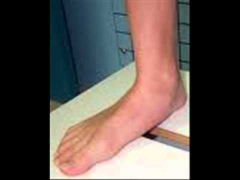
It is rare to find someone who walks with both feet in perfect alignment. A foot that is flat or has a very high arch does not necessarily require treatment unless there is pain. Often we walk on the inside or outsides of our feet, or with our toes or heels rotated inward or outward more than they should be. These atypical walking patterns can cause many problems not just with the feet but also for the whole body as the alignment of the feet sets the foundation for the whole body?s alignment. One of the most common effects of improper alignment is known as flatfoot, or pes planus.
Causes
Over-pronation is a common biomechanical problem that occurs when the arches collapse while walking or standing. This condition hampers our natural walking pattern, causing an imbalance, and leading to wear and tear in other parts of the body with every step we take. Whether you suffer from over-pronation like most of the population, or you have a true flat foot, in both cases your poor walking pattern may contribute to a range of different complaints. As we age, poor aligment of the feet causes very common conditions such as heel pain or knee pain. Over-pronation has different causes. Obesity, pregnancy, age or repetitive pounding on a hard surface can weaken the arch, leading to over-pronation. Over-pronation is also very common with athletes, especially runners, who most of them nowadays use orthotics inside their shoes.
Symptoms
People will have a very heavily dropped arch and it won?t affect them at all and people will have it slightly dropped and it could cause fierce problems. It could cause things like plantar fasciitis, it could cause heel spurs, desperate ball-of-the-foot pressure, or pressure on the big toe known as the hallux which causes discomfort in the foot. It will create problems upwards to the knees, hips and the back once you?re out of line.
Diagnosis
You can always give yourself the ?wet test? described above to see whether you have flat feet. Most people who do not notice their flat feet or have no pain associated with them do not think to see a foot doctor. Flat feet can lead to additional problems such as stiffness or pain, however, especially if the condition appears out of nowhere. If you think you may have flat feet, you should seek medical attention to ensure there are no additional issues to worry about. Your doctor will be able to diagnose you with a number of tests. For example, he or she may have you walk around, stand still, or stand on your tiptoes while you are being examined. Your doctor may also examine your foot?s shape and functionality. It?s important to let your foot doctor know about your medical and family history. In some cases, your doctor may order imaging tests such as x-rays or an MRI (magnetic resonance imaging) to determine a cause of your flat foot. If tarsal coalition is suspected in children, a CT scan is often ordered.
Non Surgical Treatment
Treatment for flat feet and fallen arches depends on the severity and cause of the problem. If flat feet cause no pain or other difficulties, then treatment is probably not needed. In other cases, your doctor may suggest one or more of these treatments. Rest and ice to relieve pain and reduce swelling, stretching exercises, pain relief medications, such as nonsteroidal anti-inflammatories, physical therapy, orthotic devices, shoe modifications, braces, or casts, injected medications to reduce inflammation, such as corticosteroids. If pain or foot damage is severe, your doctor may recommend surgery.
Surgical Treatment

Fallen arches may occur with deformities of the foot bones. Tarsal coalition is a congenital condition in which the bones of the foot do not separate from one another during development in the womb. A child with tarsal coalition exhibits a rigid flat foot, which can be painful, notes the patient information website eOrthopod. Surgery may prove necessary to separate the bones. Other foot and ankle conditions that cause fallen arches may also require surgery if noninvasive treatments fail to alleviate pain and restore normal function.
It is rare to find someone who walks with both feet in perfect alignment. A foot that is flat or has a very high arch does not necessarily require treatment unless there is pain. Often we walk on the inside or outsides of our feet, or with our toes or heels rotated inward or outward more than they should be. These atypical walking patterns can cause many problems not just with the feet but also for the whole body as the alignment of the feet sets the foundation for the whole body?s alignment. One of the most common effects of improper alignment is known as flatfoot, or pes planus.
Causes
Over-pronation is a common biomechanical problem that occurs when the arches collapse while walking or standing. This condition hampers our natural walking pattern, causing an imbalance, and leading to wear and tear in other parts of the body with every step we take. Whether you suffer from over-pronation like most of the population, or you have a true flat foot, in both cases your poor walking pattern may contribute to a range of different complaints. As we age, poor aligment of the feet causes very common conditions such as heel pain or knee pain. Over-pronation has different causes. Obesity, pregnancy, age or repetitive pounding on a hard surface can weaken the arch, leading to over-pronation. Over-pronation is also very common with athletes, especially runners, who most of them nowadays use orthotics inside their shoes.
Symptoms
People will have a very heavily dropped arch and it won?t affect them at all and people will have it slightly dropped and it could cause fierce problems. It could cause things like plantar fasciitis, it could cause heel spurs, desperate ball-of-the-foot pressure, or pressure on the big toe known as the hallux which causes discomfort in the foot. It will create problems upwards to the knees, hips and the back once you?re out of line.
Diagnosis
You can always give yourself the ?wet test? described above to see whether you have flat feet. Most people who do not notice their flat feet or have no pain associated with them do not think to see a foot doctor. Flat feet can lead to additional problems such as stiffness or pain, however, especially if the condition appears out of nowhere. If you think you may have flat feet, you should seek medical attention to ensure there are no additional issues to worry about. Your doctor will be able to diagnose you with a number of tests. For example, he or she may have you walk around, stand still, or stand on your tiptoes while you are being examined. Your doctor may also examine your foot?s shape and functionality. It?s important to let your foot doctor know about your medical and family history. In some cases, your doctor may order imaging tests such as x-rays or an MRI (magnetic resonance imaging) to determine a cause of your flat foot. If tarsal coalition is suspected in children, a CT scan is often ordered.
Non Surgical Treatment
Treatment for flat feet and fallen arches depends on the severity and cause of the problem. If flat feet cause no pain or other difficulties, then treatment is probably not needed. In other cases, your doctor may suggest one or more of these treatments. Rest and ice to relieve pain and reduce swelling, stretching exercises, pain relief medications, such as nonsteroidal anti-inflammatories, physical therapy, orthotic devices, shoe modifications, braces, or casts, injected medications to reduce inflammation, such as corticosteroids. If pain or foot damage is severe, your doctor may recommend surgery.
Surgical Treatment
Fallen arches may occur with deformities of the foot bones. Tarsal coalition is a congenital condition in which the bones of the foot do not separate from one another during development in the womb. A child with tarsal coalition exhibits a rigid flat foot, which can be painful, notes the patient information website eOrthopod. Surgery may prove necessary to separate the bones. Other foot and ankle conditions that cause fallen arches may also require surgery if noninvasive treatments fail to alleviate pain and restore normal function.
What May Cause Pain In The Arch ?
Overview
Plantar fasciitis is a common and often persistent kind of repetitive strain injury afflicting runners, walkers and hikers, and nearly anyone who stands for a living, cashiers, for instance. It causes mainly foot arch pain and/or heel pain. Morning foot pain is a signature symptom. Plantar fasciitis is not the same thing as heel spurs and flat feet, but they are related and often confused. Most people recover from plantar fasciitis with a little rest, arch support (regular shoe inserts or just comfy shoes), and stretching, but not everyone. Severe cases can stop you in your tracks, undermine your fitness and general health, and drag on for years.

Causes
Plantar fasciitis, another sports injury detailed on this website, is regularly the cause of foot arch pain or strain. This can arise due to faulty biomechanics in your feet, which alone can also provoke foot arch pains. The most prominent biomechanical difficulties are flat feet and high arches. With flat feet (or overpronation) the arches appear to be almost flattened, causing unevenness by forcing the feet roll inwards in order to maintain balance and support the body's weight. This places inordinate pressure on the plantar fascia and arches. If by contrast you have high arches (instep), the ankle can roll outwards, again causing undue strain on the arches. Too much of this strain can lead to stretching of the plantar fascia and pain in the arches. Other causes include overstretching or otherwise pressuring the arches, for example by exercising with fatigued leg muscles which leave the feet with excessive work to do. You are also particularly at risk if in your 40s or 50s and commencing an intense program of training after a long period of inactivity.
Symptoms
Persistant pain and selling under the ball of the foot and extending towards the toes (most commonly the 2nd). Some swelling may be disable on the top of the foot along with redness. Often a sensation of 'walking on the bones for the foot' will be described, and there is a positive Lachman's test. Often a tear will result in the toes splaying (daylight sign) and clawing.
Diagnosis
A patient is asked to step with full body weight on the symptomatic foot, keeping the unaffected foot off the ground. The patient is then instructed to "raise up on the tip toes" of the affected foot. If the posterior tibial tendon has been attenuated or ruptured, the patient will be unable to lift the heel off the floor and rise onto the toes. In less severe cases, the patient will be able to rise on the toes, but the heel will not be noted to invert as it normally does when we rise onto the toes. X-rays can be helpful but are not diagnostic of the adult acquired flatfoot. Both feet, the symptomatic and asymptomatic - will demonstrate a flatfoot deformity on x-ray. Careful observation may show a greater severity of deformity on the affected side.
Non Surgical Treatment
This is a common foot condition that can be easily treated. If you suffer from arch pain avoid high-heeled shoes whenever possible. Try to choose footwear with a reasonable heel, soft leather uppers, shock absorbing soles and removable foot insoles. When the arch pain is pronation related (flat feet), an orthotic designed with a medial heel post and proper arch support is recommended for treating the pain. This type of orthotic will control over-pronation, support the arch and provide the necessary relief. If the problem persists, consult your foot doctor.

Surgical Treatment
With flat feet, there is a tendon on the inside of the foot than can often become weakened, injured, split and/or ruptured. This tendon, is called the posterior tibial tendon, and is the main arch supporting tendon. Obviously damage to this tendon can cause collapse of the arch. Some people have genetically inefficient tendon, and tends to be the case in younger people. In mild cases, such as tendon splits, the posterior tibial tendon can be repaired to restore its strength. Acute incontinuity of the tendon can be primarily repaired. Often the posterior tibial tendon is augmented with a tendon transfer of an adjacent tendon to provide both strength and continuity. In any tendon repair, advanced or retensioning of the tendon is performed. In most flat foot surgery a tendon augmentation is often combined with other boney procedures to restore structure and balance to the foot.
Stretching Exercises
People with flexible feet who develop fallen arches may benefit from foot strengthening exercises, notes the Nicholas Institute of Sports Medicine and Athletic Trauma. Standing on a towel in bare feet and grasping the material with the toes is an easy foot-strengthening exercise that can be done at home. Standing on one leg while arching and releasing the foot may also prove useful. Doctors may prescribe gentle stretching exercises for the foot and ankle tendons.
Plantar fasciitis is a common and often persistent kind of repetitive strain injury afflicting runners, walkers and hikers, and nearly anyone who stands for a living, cashiers, for instance. It causes mainly foot arch pain and/or heel pain. Morning foot pain is a signature symptom. Plantar fasciitis is not the same thing as heel spurs and flat feet, but they are related and often confused. Most people recover from plantar fasciitis with a little rest, arch support (regular shoe inserts or just comfy shoes), and stretching, but not everyone. Severe cases can stop you in your tracks, undermine your fitness and general health, and drag on for years.
Causes
Plantar fasciitis, another sports injury detailed on this website, is regularly the cause of foot arch pain or strain. This can arise due to faulty biomechanics in your feet, which alone can also provoke foot arch pains. The most prominent biomechanical difficulties are flat feet and high arches. With flat feet (or overpronation) the arches appear to be almost flattened, causing unevenness by forcing the feet roll inwards in order to maintain balance and support the body's weight. This places inordinate pressure on the plantar fascia and arches. If by contrast you have high arches (instep), the ankle can roll outwards, again causing undue strain on the arches. Too much of this strain can lead to stretching of the plantar fascia and pain in the arches. Other causes include overstretching or otherwise pressuring the arches, for example by exercising with fatigued leg muscles which leave the feet with excessive work to do. You are also particularly at risk if in your 40s or 50s and commencing an intense program of training after a long period of inactivity.
Symptoms
Persistant pain and selling under the ball of the foot and extending towards the toes (most commonly the 2nd). Some swelling may be disable on the top of the foot along with redness. Often a sensation of 'walking on the bones for the foot' will be described, and there is a positive Lachman's test. Often a tear will result in the toes splaying (daylight sign) and clawing.
Diagnosis
A patient is asked to step with full body weight on the symptomatic foot, keeping the unaffected foot off the ground. The patient is then instructed to "raise up on the tip toes" of the affected foot. If the posterior tibial tendon has been attenuated or ruptured, the patient will be unable to lift the heel off the floor and rise onto the toes. In less severe cases, the patient will be able to rise on the toes, but the heel will not be noted to invert as it normally does when we rise onto the toes. X-rays can be helpful but are not diagnostic of the adult acquired flatfoot. Both feet, the symptomatic and asymptomatic - will demonstrate a flatfoot deformity on x-ray. Careful observation may show a greater severity of deformity on the affected side.
Non Surgical Treatment
This is a common foot condition that can be easily treated. If you suffer from arch pain avoid high-heeled shoes whenever possible. Try to choose footwear with a reasonable heel, soft leather uppers, shock absorbing soles and removable foot insoles. When the arch pain is pronation related (flat feet), an orthotic designed with a medial heel post and proper arch support is recommended for treating the pain. This type of orthotic will control over-pronation, support the arch and provide the necessary relief. If the problem persists, consult your foot doctor.
Surgical Treatment
With flat feet, there is a tendon on the inside of the foot than can often become weakened, injured, split and/or ruptured. This tendon, is called the posterior tibial tendon, and is the main arch supporting tendon. Obviously damage to this tendon can cause collapse of the arch. Some people have genetically inefficient tendon, and tends to be the case in younger people. In mild cases, such as tendon splits, the posterior tibial tendon can be repaired to restore its strength. Acute incontinuity of the tendon can be primarily repaired. Often the posterior tibial tendon is augmented with a tendon transfer of an adjacent tendon to provide both strength and continuity. In any tendon repair, advanced or retensioning of the tendon is performed. In most flat foot surgery a tendon augmentation is often combined with other boney procedures to restore structure and balance to the foot.
Stretching Exercises
People with flexible feet who develop fallen arches may benefit from foot strengthening exercises, notes the Nicholas Institute of Sports Medicine and Athletic Trauma. Standing on a towel in bare feet and grasping the material with the toes is an easy foot-strengthening exercise that can be done at home. Standing on one leg while arching and releasing the foot may also prove useful. Doctors may prescribe gentle stretching exercises for the foot and ankle tendons.
Why Do I Get Bunions?
Overview
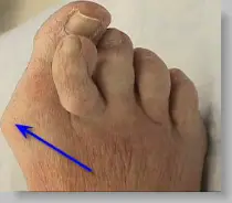
Bunions, referred to in the medical community as Hallux Valgus, are one of the most common forefoot problems. A bunion is a prominent bump on the inside of the foot around the big toe joint. This bump is actually a bone protruding towards the inside of the foot. With the continued movement of the big toe towards the smaller toes, it is common to find the big toe resting under or over the second toe. This causes a common forefoot condition called overlapping toes. Some of the symptoms of bunions include inflammation, swelling, and soreness on the side surface of the big toe. The discomfort commonly causes a patient to walk improperly. Another type of bunion which some individuals experience is called a Tailor's Bunion, also known as a Bunionette. This forms on the outside of the foot towards the joint at the little toe. It is a smaller bump that forms due to the little toe moving inwards, towards the big toe.
Causes
The commonest cause of bunions is prolonged wearing of poorly designed shoes such as the narrow high heels that women wear. This is one of the reasons why bunions are much more common in women than in men. There is also a hereditary component to bunions in that many times we will see a grandmother, mother and daughter all with various stages of bunions. 38% of women in the United States wear shoes that are too small and 55% of women have some degree of bunion formation. Bunions are 9 times more common in women than they are in men.
Symptoms
Bunions are readily apparent, you can see the prominence at the base of the big toe or side of the foot. However, to fully evaluate your condition, the Podiatrist may take x-rays to determine the degree of the deformity and assess the changes that have occurred. Because bunions are progressive, they don't go away, and will usually get worse over time. But not all cases are alike, some bunions progress more rapidly than others. There is no clear-cut way to predict how fast a bunion will get worse. The severity of the bunion and the symptoms you have will help determine what treatment is recommended for you.
Diagnosis
The doctor considers a bunion as a possible diagnosis when noting the symptoms described above. The anatomy of the foot, including joint and foot function, is assessed during the examination. Radiographs (X-ray films) of the foot can be helpful to determine the integrity of the joints of the foot and to screen for underlying conditions, such as arthritis or gout. X-ray films are an excellent method of calculating the alignment of the toes when taken in a standing position.
Non Surgical Treatment
The treatment of bunions should be individualized because the degree of deformity is not always consistent with the degree of pain. The most important first step in the treatment (and prevention) of bunions is to wear properly fitted shoes, with a low heel and adequate room in the toe area. Further treatment may include relative rest and icing to decrease pain around the MTP joint, medications to reduce inflammation and pain, stretching and strengthening exercises and shoe orthotics. If the above measures are not successful, surgery may be required.

Surgical Treatment
Conservative bunion treatment methods help to reduce the symptoms but they cannot undo changes in the bone to fully correct the condition. If the pain becomes severe and it starts to affect your daily life, then foot bunion surgery may be the best option to correct a hallux abducto valgus. There are a number of different surgical options, depending on the severity of the bony deformity. The two most common types of surgery are Osteotomy. This is the most common type pf surgery and involves removing part of the bony lump and realigning the toes. It is also known as a bunionectomy or exostectomy. The ligaments around the big toe may also need realigning and this will be done at the same time if necessary. Fusion.This is where the joint at the base of the big toe (metatarsophalangeal joint) is fused together, known as an arthrodesis. This is only indicated in severe cases or if other treatments have failed as it severely limits the movement of the big toe.
Prevention
Shoes that possess tapering toe boxes should be avoided if you have a bunion, as narrow toe boxes will hasten the progression of your bunion deformity. In some cases, conservative measures, including switching to appropriate footwear, may not have the desired effect, and your podiatrist may recommend for you a surgical procedure known as a bunionectomy.
Bunions, referred to in the medical community as Hallux Valgus, are one of the most common forefoot problems. A bunion is a prominent bump on the inside of the foot around the big toe joint. This bump is actually a bone protruding towards the inside of the foot. With the continued movement of the big toe towards the smaller toes, it is common to find the big toe resting under or over the second toe. This causes a common forefoot condition called overlapping toes. Some of the symptoms of bunions include inflammation, swelling, and soreness on the side surface of the big toe. The discomfort commonly causes a patient to walk improperly. Another type of bunion which some individuals experience is called a Tailor's Bunion, also known as a Bunionette. This forms on the outside of the foot towards the joint at the little toe. It is a smaller bump that forms due to the little toe moving inwards, towards the big toe.
Causes
The commonest cause of bunions is prolonged wearing of poorly designed shoes such as the narrow high heels that women wear. This is one of the reasons why bunions are much more common in women than in men. There is also a hereditary component to bunions in that many times we will see a grandmother, mother and daughter all with various stages of bunions. 38% of women in the United States wear shoes that are too small and 55% of women have some degree of bunion formation. Bunions are 9 times more common in women than they are in men.
Symptoms
Bunions are readily apparent, you can see the prominence at the base of the big toe or side of the foot. However, to fully evaluate your condition, the Podiatrist may take x-rays to determine the degree of the deformity and assess the changes that have occurred. Because bunions are progressive, they don't go away, and will usually get worse over time. But not all cases are alike, some bunions progress more rapidly than others. There is no clear-cut way to predict how fast a bunion will get worse. The severity of the bunion and the symptoms you have will help determine what treatment is recommended for you.
Diagnosis
The doctor considers a bunion as a possible diagnosis when noting the symptoms described above. The anatomy of the foot, including joint and foot function, is assessed during the examination. Radiographs (X-ray films) of the foot can be helpful to determine the integrity of the joints of the foot and to screen for underlying conditions, such as arthritis or gout. X-ray films are an excellent method of calculating the alignment of the toes when taken in a standing position.
Non Surgical Treatment
The treatment of bunions should be individualized because the degree of deformity is not always consistent with the degree of pain. The most important first step in the treatment (and prevention) of bunions is to wear properly fitted shoes, with a low heel and adequate room in the toe area. Further treatment may include relative rest and icing to decrease pain around the MTP joint, medications to reduce inflammation and pain, stretching and strengthening exercises and shoe orthotics. If the above measures are not successful, surgery may be required.
Surgical Treatment
Conservative bunion treatment methods help to reduce the symptoms but they cannot undo changes in the bone to fully correct the condition. If the pain becomes severe and it starts to affect your daily life, then foot bunion surgery may be the best option to correct a hallux abducto valgus. There are a number of different surgical options, depending on the severity of the bony deformity. The two most common types of surgery are Osteotomy. This is the most common type pf surgery and involves removing part of the bony lump and realigning the toes. It is also known as a bunionectomy or exostectomy. The ligaments around the big toe may also need realigning and this will be done at the same time if necessary. Fusion.This is where the joint at the base of the big toe (metatarsophalangeal joint) is fused together, known as an arthrodesis. This is only indicated in severe cases or if other treatments have failed as it severely limits the movement of the big toe.
Prevention
Shoes that possess tapering toe boxes should be avoided if you have a bunion, as narrow toe boxes will hasten the progression of your bunion deformity. In some cases, conservative measures, including switching to appropriate footwear, may not have the desired effect, and your podiatrist may recommend for you a surgical procedure known as a bunionectomy.